Monday, October 24, 2016

Electrical and Mechanical Properties of Heart (revision)

Before understanding how ECG works:
Electrical Properties of heart:
1. Autorhythmicity
The ability to generate action potentials without external stimulus
•spontaneous pre-potential (pacemaker potential) followed by action potential.
•Pacemaker tissue (sino atrial node, atrio ventricular node, atrio ventricular bundle and purkinje fibers) can initiate repetitive action potentials.
•Pacemaker tissue is characterized by unstable resting membrane potential because of its continuous change in membrane permeability.


Image result for action potential of pacemaker tissue


Pre-potential:
Slow decrease in K+ efflux while permeability of other ions remain constant (through ‘h’ channels and transient ‘T’ Ca2+ channels).

Depolarization:
Ca2+ influx through long lasting Ca2+ channels ( ‘L’ )

Repolarization:
K+ efflux.

Rate of Impulse (per min):
SA node--70-100
AV node--40-60
Atrial and atrial pathway--20-40
Purkinje fibers--20-40

2. Excitability:
Cardiac muscle is excitable tissue
-->it forms a wave of depolarization in response to a stimulus(generation of AP)
Image result for cardiac muscle action potential
0: Rapid depolarization.
Opening of Na+ channels causes increase in Na+ influx.

1: Rapid initial fall in membrane potential (initial rapid repolarization)
Inactivation of Na+ channels.

2: Plateau phase.
Slow opening of Ca2+ channels causes Ca2+ influx.

3: A rapid fall in membrane potential (repolarization phase)
Opening of K+ channels causes rapid K+ efflux.

4: Polarised state.
Ionic composition is restored by activation of Na+K+ATPase pump

3. Conductivity
the ability of cardiac muscle fibers to conduct the cardiac impulses that are initiated in the SA node.

  • Impulse from SA node spread quickly to AV node via atrial (internodal) pathway.
  • Time delay (0.1 s) occurs as impulses pass through AV node.
  • Impulse conduction increases as spread to Purkinje fibers at a velocity of 4 m/s.
  • Ventricular contraction begins 0.1-0.2 s after contraction of the atria. (important for ventricular filling phase)
  • LBB starts before RBB, as LV wall is thicker so the impulse needs more enough time to reach. Accordingly both ventricles will contract together.
******************************************************

Electrical activity of the heart:
1. Depolarization and origin of cardiac impulse at the SA node. (if SA node doesnt work, AV will take place of being the pacemaker)

2. Conduction of the impulse to all parts of the atria through atrial muscle (there may be some special pathways that conduct impulse fast ie. have a higher conduction velocity). The impulse also spreads to the AV node along the atrial muscle. (Atrial fibrillation occurs when the atrial ms contract independently and continuously bombard AV node with depolarizing waves of varying strength, and depolarization spreads at irregular intervals down the Bundle of His)

3. The impulse will not pass from atrial muscle to ventricles directly as they are not in contact (fibrous tissue separates them). (thus if AV node is blocked, and impulse from SA node couldnt reach ventricle, ventricular muscle will initiate its own impulse as it also has autorhythmicity, eg. seen as AV dissociation on ECG)

4. AV node has a low conduction velocity and thus the impulse takes sometime to travel across it. It eventually travels across it and reaches the upper end of the bundle of His.

5. Bundle of His has a high conduction velocity. It conducts the impulse through its branches to the Purkinje fibre network just under the endocardium of the ventricles. (In a normal conditionn where impulse is able to be transmitted from AV node to Bundle of His, impulse created by ventricular muscle itself doesnt cause muscle contraction as the impulse hits the absolute refractory period--all/none law of the action potential of cardiac muscle that is stimulated by normal conduction system's impulse)
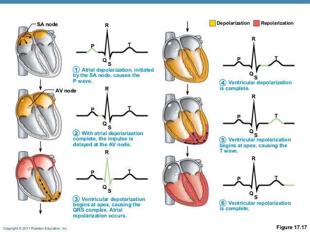
Image result for sequence of depolarization in heart

*atrial repolarization not seen on ECG as it was masked by the stronger ventricle depolarization

******************************************************
Mechanical Properties of the heart:
1. Contractility
---when an action potential occurs in the muscle it responds by contraction.
During excitation-contraction coupling the ER releases some Ca++ ,but to have sufficient calcium for contraction there must be influx through the sarcolemma. Hence the importance of the plateau phase of the action potential.

  • For cardiac muscle, the amount of stretch of the muscle before a contraction has special significance and is called preload (Starling Law).
  • Protected from tetanus because of its long ARP
  • No recruitment of cardiac muscle fibers.
  • Strength of contraction of cardiac muscle depends to a great extent on the concentration of calcium ions in the ECF
2. All or None Law

  • Action potential of cardiac muscle is an all or none law response to a stimulus.
  • If the stimulus is sub-threshold (inadequate), no action potential is produced.
  • If the stimulus reaches threshold, a full-fledged action potential is produced.
  • Further increase in the intensity of a stimulus produces no increment in the amplitude of AP.
Absolute refractory period:
  • The excitability of cardiac muscle is completely lost during this period, i.e. doesn’t respond to 2nd stimulus.
  • Occupies the whole period of systole.
Relative refractory period:
  • The excitability of cardiac muscle is partially recovered during this period, i.e. stronger stimuli than normal are required to excite the muscle.
  • Occupies the time of diastole.
  • Can be affected by the heart rate, temperature, vagal stimulation, sympathetic stimulation & drugs.
******************************************************
Factors affecting heart rate:
1. Sympathetic stimulation

Act via β1-adrenergic receptors -->increase cAMP-->Increases heart rate by opening of Ca2+ channels--> increase Ca2+ influx-->increase rapidity of the depolarization phase--> increase heart rate (positive ‘chronotropic’ effect)

2. Vagal stimulation
Acts via M2 muscarinic receptors -->decreasing cAMP-->Decreases heart rate by opening of K+ channels and slows the opening of Ca2+ channels --> increase K+ efflux of nodal tissues -->hyperpolarization --> decrease slope of pre-potentialp--> decrease firing rate--> decrease heart rate (negative ‘chronotropic’ effect)

3. Temperature
Arise in body temperature by 1 °C increases the heat rate by 10 beats/minute.
The rise in body temperature increase the heart rate by increasing the permeability of the membrane to Ca++ during the pacemaker potential.

4. Sinus arrhythmia
Variation in R-R interval during deep inspiration.
During deep inspiration, lung stretch--> inhibits cardio-inhibitory center-->decreases tonic vagal discharge -->increases HR.

5. Ion concentration in ECF
K +
-the early effect of mild hyperkalemia on myocyte function is to increase myocyte excitability by shifting the resting membrane potential to a less negative value and thus closer to threshold potential; but as potassium levels continue to rise, myocyte depression occurs and Vmax continues to decrease.

A plasma potassium of >6.5mmol/L (normal 3.5-5.5) is an emergency, myocardial hyperexcitability--> ventricular fibrillation--> cardiac arrest

5.5-6.0 mEq/L - Mild


6.1-7.0 mEq/L - Moderate


7.0 mEq/L and greater - Severe


Sx: fast irregular pulse, chest pain, weakness, palpitation, and light-headedness

ECG: Tall tented T wave, small p wave, wide QRS complex


Causes:

Decreased or impaired potassium excretion - As observed with acute or chronic renal failure (most common), potassium-sparing diuretics, urinary obstruction, sickle cell disease, Addison disease, and systemic lupus erythematosus (SLE)


Additions of potassium into extracellular space - As observed with potassium supplements (eg, PO/IV potassium, salt substitutes), rhabdomyolysis, and hemolysis (eg, blood transfusions, burns, tumor lysis)


Transmembrane shifts (ie, shifting potassium from the intracellular to extracellular space) - As observed with acidosis and medication effects (eg, acute digitalis toxicity, beta-blockers, succinylcholine)


Fictitious or pseudohyperkalemia - As observed with improper blood collection (eg, ischemic blood draw from venipuncture technique), laboratory error, leukocytosis, and thrombocytosis


Management:
Urgent:
  • Stabilize Cardiac membrane with 10ml 10% calcium gluconate
  • drive K+ into cell with 10 units actrapid in 50ml 20% glucose
Non-urgent: (K+ not >6.5, no myocardial hyperexcitability)
  • Treat underlying causes.
  • Polystyrene sulfonate resin 15g/8h PO
Ca2+ ion in ECF
  • Increase in Ca2+ concentration --> increase cardiac contractility
  • Decrease in Ca2+ concentration --> decrease cardiac contractility

6. Blood flow
Insufficient blood flow--> decreased oxygen and nutrient supply--> decreased heart conductivity and metabolism

7. Drug
Sympathomimetic drug--> increased HR 
parasympathomimetic drug--> reduce HR

Tuesday, September 20, 2016

Complete/Incomplete Spinal Cord injury, Neurological Examination and Asia Chart

Spinal shock can occur and last for about 24-72 hours after spinal cord injury and a/w complete motor and sensory loss, hypotension and bradycardia. (monitor Bp with vasopressor to prevent fluid overload)

Bulbocarvenosus reflex: 
If present, Squeezing of penis/clitoris/pulling of Foley catheter would cause contraction of anal sphincter muscle.


The reflex is spinal mediated and involves S2-S4.

Presence of reflex indicates ends of spinal shock.
(Sometimes absence of reflex could be due to cauda equina syndrome instead of spinal shock)

Sacral Sparing Test was done after ends of spinal shock to determine complete or incomplete spinal injury:
1. flexion of big toe present
2. anal reflex present
3. perianal sensation present (S3-5)

Sacral sparing indicates incomplete spinal cord injury

(pinprick sensation predicts better prognosis of motor function: as lateral spinothalamic is near to lateral corticospinal tract)

spinal cord anatomy:


TYPES OF INJURY:
Central Cord Syndrome


Anterior Cord Syndrome


Brown Sequad


Posterior Cord Syndrome


Determine Level of Spinal Cord Injury/Lesion by using Asia Chart


Difference in level of nerve exit:


Cervical nerve exit above vertebra level, example C4 nerve exit above C4 vertebra and below C3 vertebra

Thoracic nerve exit below vertebra level, example T4 nerve exit below T4 vertebra and above T5 vertebra

Loss of sensation at the dermatomal area and power at myotomal area at C4 spinal cord level would indicates injury at C3 vertebral level. Likewise a radiograph of C3 vertebra injury is expecting sensory and motor impairment to C4 spinal cord level.

In PID (Prolapsed Intervertebral Disc), the location of herniated disc in after level of conus medullaris (L1 vertebra) would determine different type of lesion.

Example, centrally herniated disc would affect nerve of the next vertebral level, lateral herniated disc would affect nerve of the same vertebral level. (Refer diagram below)

ASIA chart impairement score:
Motor power determine grade of impairment:


Summary:

In acute spinal injury (emergency case):
-check patient vitals, bulbocarvenosus reflex to rule out spinal shock, if shock present, manage the shock
-then sacral sparing test to determine complete or incomplete injury
-then neurological examination to determine type and level of injury

1. Neurological Examination (usually exam will specify either upper limb/lower limb):
a. Sensory, dermatome (Light touch, Pin Prick)
b. Motor, (Tone, Power, Reflex)
*always compare both side

2. Determine Type of  Injury:
a. Pure sensory loss
b. Pure motor weakness
c. Combined motor and sensory loss
d. Unilateral/Bilateral

3. Determine Etiology:
Acute: Fracture(burst) 
Gradual: PID, spinal stenosis

4. Confirm diagnosis with special test
eg. Straight leg raising test in PID, perianal sensation to rule out cauda equina syndrome (usually caused by large midline disc herniation or extrusion, emergency and need surgical decompression if patient had urinary retention)

Sunday, August 14, 2016

Remedial O&G Group 1 teaching with Dr. Rahimah [note to myself]

Dr. Rahimah stressed on knowing definition of gravida, parity and knowing how to count LMP and EDD. In case patient is USOD, we can give roughly estimated LMP by subtracting 40 weeks from REDD.

eg if this is patient 5th pregnancy, she had a molar, ectopic, a miscarriage and an IUD so it should be?

G5P1+3 (where there is 1 molar, 1 ectopic and 1 miscarriage)

In a case of SGA/IUGR, I put 'or' because a SGA case(suspected constitutionally small because of smaller mother's size with previous hx of small fetus) must be managed as TRO IUGR as if causes failed to be identified and patient was discharged and if a IUGR was not detected and managed, it will lead to IUD.

3 important factors lead to IUGR:
1. Placental insufficiency, which could be caused by
-pre-eclampsia (usually early onset which start to affect during formation of placenta)
-chronic medical illness which affect placenta formation
-smoking
-autoimmune disease such as SLE
-placenta previa, as placenta attach to lower segment of uterus with thinner myometrium--> less blood supply--> less nutrients--> baby smaller
(thats why we opt for lower segment c-sec as lesser blood supply there)

2. fetal infection
rule out TORCHES

3. fetal anomalies
oligohydramnios suspect renal agenesis

symmetrical IUGR: early onset, caused by Chromosomal anomalies, infection
asymmetrical IUGR, late onset, caused by placental insufficiency, thus have head-sparing

GDM will cause fetal macrosomia
however pre-existing DM in mother more likely --> congenital anomalies--> fetal microsomia

as this patient has a previous hx of small baby, ask if there is oligohydramnio in the previous pregnancy, chances are the patient has intrauterine infection in the previous pregnancy which continue to the next...

find out about percentile on growth chart for SGA, IUGR

if at district hospital, need to refer to tertiary centre if there are two abnormal plotted growth charts
usually the gap between 2 serial scans is about 1 week

however normal growth scan is 2 weeks

1 previous scar is already a criteria for admission

If Doppler U/S is abnormal, need to do CTG daily

If leaking of liquor, need to do U/S every 1w or 3d

HOPI for IUGR must rule out any CX such as
abnormal CTG, reduced fetal movement,doppler u/s findings, leking of liquor any PV discharge

Also diet hx to assess whether mother malnourish

VE assess Bishop score, if cervix favourable, deliver
if not, do induction, however previous scar, so do not do Induction as risk of uterine rupture...
if fetal distress, abnormal CTG, doppler, then c-sec
if no fetal distress, opt for vaginal delivery

Plan and management:
1. monitoring
2. timing of delivery
3. mode of delivery

investigations:
1. Blood: anemia, TWC, torches screening, group screen hold, High vaginal swab if leaking of liquor
2. U/S doppler: fetal BPP, placenta (any redding calcification), head and abd circumference, any fetal anomalies such as renal agenesis
3. CTG daily, fetal heart monitoring,

what to revise:
growth chart
causes and mx of IUGR
fetal BPP
torches
redding calcification


Tuesday, August 9, 2016

Remedial O&G CT3 & 6 [note to myself]

CT 6 teaching with Dr. Hoo

Pt is a 32y/o lady G2P1 came in with Placenta Previa Type III Posterior for observation at 34w + 5d.
She had previous hx of Placenta Previa Type IV in her first pregnancy and had done elective Cesarean section at 2013 which is 3 years ago. She also had a past surgical history of cystectomy at 2012 which is 4 years ago. She denied any APH such as PV bleed or abd pain. The dx was made based on ultrasound findings at 30w.

In this pt, the risk factor for her recurrent PP in second pregnancy is previous C-sec.
To clerk history of a previous C-sec, must exclude cx such as any blood transfusion? any injury to bladder, bowel, ureters? any post-partum hemorrhage? any wound infection? endometritis? UTI?
Importance is to detect any presence of placenta accreta in her next pregnancy...as C-sec is a risk.

C-sec is also planned for her second pregnancy.
Must know how to plan a C-sec,
Inform and get consent,
Large bore IV access, catheterize bladder,

Pre-op: Take blood for FBC (ensure Hb level >10g/dl), PT/PTT, GXM (4-6units of blood), urea/creatinine.
consider ECG/CHEST XRAY

Neutralize gastric contents to prevent aspiration pneumonia if GA is used.
Elective C-sec, NBM + Ranitidine

Emergency C-sec: sodium citrate and metoclopromide

Thromboprophylaxis
low-risk: early mobilization, hydration
moderate: IV heparin and TED stockings
high: IV heparin until 5-d post-op and TED stockings

Antibiotic prophylaxis
IV cefazolin. given after cord is clamped

Also don't forget to revise all the risk factors that could cause PP.

Learning Point:
C-sec cx and planning
GXM and GSH difference
PP type, risk factors, mx
Rubella=german measles, effect on fetus?
ATT to prevent emergency delivery at non sterile environment
Hepatitis B vaccine? can be given during pregnancy or not?
Primary vs secondary dysmenorrhea
Pap smear done because need to remove pre-malignant lesion if there is.
Find out about Diabetic diet and ensure GDM mother follow
Results for MOGTT 5.6-7.8

CT3 with Dr. Fauziah

Know what is abnormal progress of labour, the hour...
Know partogram, and how to detect abnormal progress by partogram and how to mx to avoid fetal distress

what can a 1st U/S tell us?
-viability,
-dating
-location of gestational sac (ectopic: abdomen, cervix, broad ligament)
-fetal number, MCDA, DCDA
-Pregnancy of unknown location need beta-hCG test
-exclude uterine abnormalities
-exlude fibroid, ovarian mass

Ovarian cyst in pregnancy, cystectomy can only be done less than 18w, more than 18w might cause abortion, so planned cystectomy only after delivery

MOGTT HOW TO DO, RESULTS, WHEN TO DO, RISK FACTOR


Sunday, August 7, 2016

Remedial O&G Fetal events during labour


AP diameter:
Suboccipitobregmatic 9.5 cephalic (well-flexed)

Transverse diameter:
Biparietal: 9.5cm
Bitemporal: 8cm


Internal rotation probably occurs as the fetal head meets the muscular sling of pelvic floor, it is often not accomplised until presenting part has reached the level of ischial spine (zero station) and is therefore engaged.

engaged= 2/5 palpable on abdomen = zero station descent at ischial spine.

OBS HX TAKING (WHY I CLERK?) [note to myself]

Obstetric hx taking learning point:

Pt identification data: to assess risk, eg age>25 must do MOGTT, eg. If you get pregnant at age 25, your risk of having a baby with Down syndrome, for example, is about 1 in 1,250, according to the National Institutes of Health. At age 40, the risk is 1 in 100. If you'll be 35 or older on your due date, you'll be offered genetic counseling.

Occupation, socioeconomic class, does the mother able to take care of the baby after C-sec at home? Does the mother BF when she needs to go to work? the home using well water? can she take water after surgery while recovering?

gravida: G, P and +
multipara will has dx of false labour if dilatation is only 1cm
if +3 indicates recurrent miscarriage, need to find out cause

LMP: naegeles rule, SOD, regular menses, no BF, no hormonal therapy
EDD, REDD, POA
(must knw how to count)
used to detect postdate and also date for IOL for cases like GDM, PIH,...
date also help to identify IUGR, SGA...
read about U/S and discrepancy

C/O what bring pt to hospital? electively admitted? referred case?
(pt came in due to contraction pain? distinguish from braxton hicks, VE see any favourable cervix using Bishop score, ask for any show/leaking of liquor)


HOPI:
must knw disease:

1. GDM/pre-existing DM, clerk MOGTT, presence of risk factors, glycosuria, sx of DM (3Ps, numbness, blurring of vision), HbA1C, family hx of GDM, any congenital anomalies in fetus, any macrosomic baby, polyhydramnios, currently admitted blood sugar profile, insulin how many unit taken, any diet control? mx: IOL? cx that may arise, macrosomic, shoulder dystocia, perineal tearing? neonatal hypoglycemia?

2. Pre-eclampsia, Bp, proteinuria, mx

3. Placenta previa, PV bleed? pain? woody hard uterus?(abruptio) grade of placenta previa, causes such as multiparity,..., mx Mcafree Regime!

4. Postdate

5. Oligohydramnios, how to see AFI

6. Abnormal lie, whether to manually betulkan lie or let it be

anemia, iron metabolism
CVD, how to terminate pregnancy

IOL indications, C-sec indication, trial of scar


HOPP
missed period? UPT? (read UPT test principle, hCG, amino acids...)
failed contraception or planned pregnancy? assess pt's desire of having baby

if not because of missed period? sx of pregnancy? vomitting, abd distension, quickening?

confirm pregnancy where?

BOOKING! READ ABOUT EACH COMPONENT OF BOOKING AND KNOW WHY WE SHOULD KNOW.

(every month till 28, every  2w  until 36, every 1w until delivery)

SUBSEQUENT FOLLOW UP? ULTRASOUND OK?

quickening? primid 18-20w, multi 16-18w

sx of pregnancy, breast engorgement, frequency, constipation, ankle edema, backache? (look for any symptomatic tx?)

Immunization
ATT (principle)
hep B (3x) last time...
Rubella secondary sch

Past OBS

consanguinous marriage?

Mode of delivery, Complication? preterm, full term? C-sec? episiotomy? poor spacing? healing scar? exclusive breastfeeding?
Recurrent miscarriage?  D&C? (uterine scar...risk of placenta previa...)

any contraception?

Menstrual hx

menorrhagia? oligomenorrhea? amenorrhea?
PCOS? subfertility?

Gynae

PCOS? Uterine fibroid? any malignancy? ectopic pregnancy? recurrent miscarriage?

Pap smear done? when? where? result?  (check how many times pap smear must be done every duration?)

PAST MEDICAL, SURGICAL, DM? HPT? HEART DISEASE? ASTHMA? TB?
uterine surgery, risk of placenta previa...

Family hx?
GDM, DM, HPT, HEART DISEASE, twins pregnancy, recurrent miscarriage, stillbirth, congenital abnormalities, gynae malignancy...

Personal and Social hx
socioeconomic status
does husband take care of family
children under care of
smoking, alcohol (husband? pt?)

Drug hx
iron, folic acid, vit b, vit c
*vit b complex contained folic acid
*obimin contain all

insulin for GDM (read back why Oral hyperglycemic Agent cannot be given)
magnesium sulphate for PIH (not sure, read back)

Prostin induction of labour, syntometrine, pitocin...(how to use)

Miseprostol, mifepristone for expulsion of POC

check for drugs use for termination of pregnancy

progesterone for maintaining pregnancy in threatened miscarriage

infection in pregnancy how to treat? common: UTI

any traditional med? any OTC drugs? any allergy?

Dietary
DM diet

Summary
AGE, GRAVIDA, PARA, POA, C/O

Remedial O&G CT1,2 [note to myself]

CT 1: GDM case teaching by Dr. Akram
Booking at least at 6w
MOGTT when, result? Insulin unit? BSP controlled? HbA1C? U/S: congenital anomalies? polyhydramnios? macrosomia?
Use SFH to compare with POG
FHR must show u're counting for 1 min
if pre-eclampsia PE must check liver tenderness, epigastric pain

CT2: Placenta Previa case teaching by Prof Adibah
quickening at 28w? impossible, check LMP wrong date/abdomen too thick

how to present:

she noticed she is pregnant when UPT is done and confirmed her pregnancy after she experienced nausea, vomitting,...

Booking was performed at....
the parameters are as followed...
u/s done at....w, and showed fetus corresponding to dates, subsequent checking was done regularly, throughout her pregnancy, there was no hypertension,...

quckening at....w

1st dose of ATT was given at....w, subsequent dose at...w

at 29w, another u/s performed as routine procedure which show placental low-lyng,...otherwise no fetal abnormalities, however, never has APH,

ask any fibroid, previous uterine surgery (risk factor)

PE for placenta previa:
1. Pain? contraction pain can cause APH in placenta previa

2. Vital signs
Bp: rule out late onset of HPT, as pt has family hx
HR: Wolff-Parkinson white syndrome...due to hyperactive thyroid

3. pallor, signs of anemia, make sure pt not in dilutional anemia, Hb enough to compensate when APH happen

4. edema, she is at risk to develop HPT, so if very gross edema, assess renal problem...

Remedial O&G CT4 [note to myself]

Clinical teaching 4 with Prof Nik

Madame NH 24y/o, G1PO with a background history of subfertility for 3 years and undergone ovulation induction for 1 year (on Clomiphene ?dose) was electively admitted for management of postdate.

DOA: 6/8/2016; DOC: on the same day as admission

Last Menstrul Period: 2/12/2015; sure of date, irregular menstruation, ovulation induction

Ovulation induction(read on infertility IDA lecture) signifies there is ovulation during the LMP so LMP is trustable.

Expected date of delivery: 9/8/2016

Revised EDD(scan done around 9th week): 31/7/2016

Prof mentioned scan done around 9th week discrepancy can be up to 2-5d, find out u/s and discrepancy

the discrepancy was 1 week and so EDD counted from LMP is acceptable. (discrepancy not more than 2 weeks; find out)

currently POA: 39w 5d (if the case is presented as a Postdate case, then do not present 39w, not tally)

HOPI:

All parameters normal, excpet on ultrasound noted cord around neck and aminiotic fluid index: 1.3cm. (learn how to grade oligohydramnios, causes and management)

Beware on small AFI, usually on PE, u may find smaller fundal height

Patient is obese class I with BMI of 30.06kgm-2. (learning point: obesity cause PCOS hence irregular menstruation and subfertility?)

Subfertility, clomiphene: must ask for dosage(mg),  how many times taken a day, duration taken (clomiphene usually 1 tab 50mg OD, given at 5th to 9th or 2nd to 6th day of menstrual cycle)

noted that BMI >30, MOGTT as screening of diabetes must be done at 12-14w, 24-28w and 32-34w,
other risks factor:


  1. Gestational diabetes previous pregnancy
  2. Obesity (BMI >30)
  3. Age > 25
  4. Presence of glycosuria in >2 occasions
  5. History of DM in first degree relatives
  6. Previous big baby > 4.0 kg
  7. Previous history of recurrent abortion or unexplained stillbirth
  8. Previous congenital anomalies
  9. Polyhydramnios

Physical examination:
rather than mention no resp distress, mention pt is breathing spontaneouly, well
only 5/5 palpable can be presented as balloptable fetal head (~floating)
Pt has high BP, must check CVS! check for radiofemoral delay, Coarctation of aorta! Treat pt as whole!
if not checking the breast, at least ask the pt whether there is presence of colostraum, which prepare pt for BF, student might miss pregnant mother who has breast cancer

Patient also has past medical hx of leukemia (unclear hx: so must ask what age diagnosed? completed chemotherapy? regularly follow up? whether cytotoxic drugs had caused the subfertility? and also a leukemic pt in the past need to have antenatal follow up in a tertiary centre! not district)

finally, presenter was asked to present CTG as there is cord around neck,

Prof's comment on how to present, this is a CTG of Madame NH, 24y/o Malay lady, G1PO, with a background hx of subfertility for 3 years and was on ovulation induction for 2 years, the CTG was taken on yesterday, 6/8/2016 2pm for a duration of _ hour, it shows that the baseline FHR was 140-150bpm, the beat to beat variability was 5-20bpm, there was presence of acceleration, no deceleration, the CTG taken is reactive. (any contraction? there was presence of regular uterine contraction with amplitude of?)

eventually, patient was given elective Caeserean section due to these following issue, subfertility, oligohydramnios and cord around neck. 

SO AGAIN, 
LEARNING POINT:
SUBFERTILITY CAUSES, CLOMIPHENE DOSAGE
PCOS
ULTRASOUND DISCREPANCY
OLIGOHYDRAMNIOS
CORD AROUND NECK
INDICATION FOR IOL/C-SEC (HOW TO CHOOSE)
HOW TO MANAGE POSTDATE IF NO CX ARISE?

EXTRA LEARNING POINT:
REVISE GDM
REVISE CVS, what may cause High Bp, RISK FOR pre-eclampsia

I asked a question why Prostin used in IOL is contraindicated in severe asthma/glaucoma?
Prof: only "severe" and its not absolute contraindication, if pt's asthma/glaucoma condition is well-controlled, Prostin can be used.


Monday, February 1, 2016

Lower Limb Peripheral Nerve Examination

Femoral nerve (L2,3,4)
motor: iliopsoas, test with hip flexion against resistance, 
           quadriceps, test with knee extension against resistance (also inspect for any wasting, check 
           knee jerk)
sensation: front of thigh, medial of leg and foot
usually caused by hematoma of illacus ( hemophilia or hip extension injuries)

Common peroneal nerve (lateral popliteal) L4,5,S1,2
motor: ant compartment (ant tibialis, extensor hallucis longus, extensor digitorum longus, peroneus tertius)
           peroneal compt (peroneus longus n brevis)
           foot ( extensor brevis)

test dorsiflexion (deep branch) and evertion (sup branch) of foot

sensation: first web of foot,dorsum of foot, front and side of leg

usually caused by if @ fibular neck, trauma (lateral ligament injury of knees or direct blow), pressure (cast/side iron of thomas splint; ganglion), ischemia ( tourniquet)
 if distal to fibular nerve, ant compartment syndrome

Tibial nerve (L4-S3)
motor: post comprt ( gastrocnemius, post tibialis, flexor hallucis longus, flexor digitorum longus)
           all the ms of sole via medial n lateral plantar n

inspect, ms wasting of sole, clawing of toe, trophic ulceration, test for power of toe flexion

sensation, sole of foot, dorsum of toe and nailbed
side of foot by sural nerve (tibial mix common peroneal)

proximal lesion of tibial nerve (wasting and loss of plantarflexion)

causes: proximal tibial fracture. post compartment syndrome, tight cast, diabetic neuropathy, tarsal tunnel syndrome


sciatic nerve (L4-S3)
motor loss: hamstring of thigh, tibial and peroneal nerve palsy
sensory loss: entire sole, foot, dorsum of foot, lateral aspect of leg, lateral half of calf, (if post cutaneous nerve of thigh is involved then back of thigh also lost sensation)
causes, usually post dislocation of hip, wound to post thigh (do not confused with sciatic palsy  2 to root involvement caused by PID)
absent ankle jerk

Lateral cutaneous n of thigh (L2,3)
maybe compressed by inguinal ligament (pain n parasthesia) or by spinal stenosis
sensory: lateral aspect of thigh
test, pressure over the nerve may give parasthesia to the thigh

neurological control of bladder, s2,3,4 supplies detrusor muscle of bladder and internal spincter

if cord transected above L2, thoracic spine fracture, voluntary control loss, still able to empty bladder 200-400ml every 2-4 hours, automatic bladder
if sacral centre damage or damage to cauda,  loss of reflex, atonic bladder

Tuesday, January 5, 2016

Bowel Preparation

 Taken from HUSM Endoscopy Unit
1 day prior to appointment
(Breakfast only)
Allow 2 piece of plain bread or plain mee and plain water. Do not take any meat, vegetables, fruits or any dairy product (produk tenusu). Please drink at least 2 litre of plain water until midnight. (If needed, take glucose drink dibenarkan sahaja jika perlu).

3pm and 7pm
Add 45ml of FLEET into a glass of plain water. Finish the dilution. This preparation(cirit-birit) works within 30 mins but may take as long as 3 hours.

12 midnight
Start fasting until colonoscopy done.

*********************************************************************************
Taken from H K.L Nurse's blog

Colclean or Fleet solution.(for patients with no renal impairment)
  • ·         Avoid eating any dietary fibers at least 3 days before the scheduled procedure. E.g.: veggies, fruits, oats, etc. (for outpatients)
  • ·         Can eat Breakfast and lunch.
  • ·         Preparation must be done the day before the Colonoscopy procedure
  • ·         45mls Colclean / Fleet + a minimum of 3 glasses of water to be given to the patient at 3.00pm. (No more meals are to be taken after the first dose.)
  • ·         Your patient will start to have Diarrhea soon afterwards.
  • ·         Another 45mls + a minimum of 3 glasses of water to be given at 6.00pm
  • ·         Patient should be encouraged to drink lots of clear fluids afterwards to avoid dehydration and given IV Drips if necessary. Your patient is going to lose a lot of fluids.
  • ·         Patient is allowed to drink clear fluids. No need NBM. (consult your Gastro M.O first)
  • ·         On the day (8.00am) of the planned procedure, call Scope room to inform the case.
  • ·         Call scope room if you are not sure about the bowel prep.


Fortrans - Poly Ethelyne Glycol (most Gastroenterologist likes to use this)
  • ·         Avoid eating any dietary fibers at least 3 days before the scheduled procedure. E.g.: veggies, fruits, oats, etc. (for outpatients)
  • ·         Can eat Breakfast and lunch.
  • ·         One sachet of Fortrans is to be diluted with one liter of clear fluid.
  • ·         3 sachet = 3liters
  • ·         One liter of diluted Fortrans is to be taken at 6.00pm
  • ·         Another liter of fortrans at 7.00pm
  • ·         And another one at 8.00pm
  • ·         All Diluted fortrans should be finished by the patient.
  • ·         Patient should be encouraged to drink lots of clear fluids afterwards to avoid dehydration and given IV Drips if necessary. Your patient is going to lose a lot of fluids.
  • ·         Patient is allowed to drink clear fluids. No need NBM. (consult your Gastro M.O first)
  • ·         On the day (8.00am) of the planned procedure, call Scope room at to inform the case.
  • ·         Call scope room if you are not sure about the bowel prep.
  • this solution is safe for patients with fluid restrictions because it does not absorb the fluid in the system.
Now, please make sure that your patient has a FUNCTIONING I.V access (branula) before you send them to the scope room.

*FLEET can't be used for renal impairment or cardiac failure patient because the sodium will cause fluid overload.