Monday, November 16, 2015

Bronchiolitis in children


INTRODUCTION
Bronchiolitis is a lower respiratory tract infection that occurs in children younger than two years old. Differential for rapid breathing and wheezing in child less than 2 years old, usually asthma only be diagnose >2 years old and with strong history of atopy. It is usually caused by a virus. The virus causes inflammation of the small airways (bronchioles) (figure 1). The inflammation partially or completely blocks the airways, which causes wheezing (a whistling sound heard as the child breathes out). This means that less oxygen enters the lungs, potentially causing a decrease in the blood level of oxygen.
Image
Bronchiolitis is a common cause of illness and is the leading cause of hospitalization in infants and young children. Treatment includes measures to ensure that the child consumes adequate fluids and is able to breathe without significant difficulty. Most children begin to improve two to five days after first developing breathing difficulties, but wheezing can last for a week or longer. Bronchiolitis can cause serious illness in some children. Infants who are very young, born early, have lung or heart disease, or have difficulty fighting infections or handling oral secretions are more likely to have severe disease with bronchiolitis. It is important to be aware of the signs and symptoms that require evaluation and treatment.
Bronchiolitis is typically caused by a virus. Respiratory syncytial virus (RSV) is the most common cause.
In tropical and semitropical climates, the seasonal outbreaks usually are associated with the rainy season. (Nov-Jan)
Virtually everyone will have been infected with RSV by the age of three years. It is common to be infected more than once. however, subsequent infections are usually milder.
Children who are older than two years typically do not develop bronchiolitis, but can be infected with RSV.  It usually causes symptoms similar to those of the common cold or mild wheezing.
Bronchiolitis usually develops following one to three days of common cold symptoms, including the following:
Nasal congestion and discharge.
A mild cough.
Fever (temperature higher than 100.4ºF or 38ºC). How to take temperature in a child?
Decreased appetite.

As the infection progresses and the lower airways are affected, other symptoms may develop, including the following:
Breathing rapidly (60 to 80 times per minute) or with mild to severe difficulty
Wheezing, which usually lasts about seven days
Persistent coughing, which may last for 14 or more days (persistent cough also may be caused by other serious illnesses that require medical attention)
Differential for persistent cough:
  • recurrent resp infection
  • post-specific resp infection (pertussis, RSV, mycoplasma)
  • asthma
  • suppurative lung disease (cystic fibrosis, ciliary dyskinesia or immune disease)
  • recurrent aspiration (GERD)
  • persistent endobronchial infection
  • inhaled foreign body
  • cigarette smoking (active or passive)
  • TB
  • Habit cough
  • airway anomalies (tracheo-bronchomalacia, tracheo-oesophageal fistula)

Difficulty feeding related to nasal congestion and rapid breathing, which can result in dehydration
assess dehydration in the child
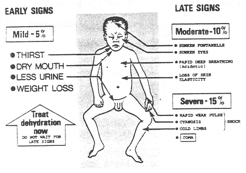
Apnea (a pause in breathing for more than 15 or 20 seconds) can be the first sign of bronchiolitis in an infant. This occurs more commonly in infants born prematurely and infants who are younger than 2 months.

Signs of severe bronchiolitis include retractions (sucking in of the skin around the ribs and the base of the throat) (figure 2), nasal flaring (when the nostrils enlarge during breathing), and grunting. The effort required to breathe faster and harder is tiring. In severe cases, a child may not be able to continue to breathe on his or her own.

Image

Low oxygen levels (called hypoxia) and blue-tinged skin (called cyanosis) can develop as the illness progresses. Cyanosis may first be noticed in the finger and toenails; ear lobes; tip of the nose, lips, or tongue; and inside of the cheek. Any of these signs or symptoms requires immediate medical evaluation.
A child who is grunting, appears to be tiring, stops breathing, or has cyanosis needs urgent medical attention. 
Contagiousness — The most common cause of bronchiolitis, respiratory syncytial virus (RSV), is transmitted through droplets that contain viral particles; these are exhaled into the air by breathing, coughing, or sneezing. These droplets can be carried on the hands, where they survive and can spread infection for several hours. If someone with RSV on his or her hands touches a child's eye, nose, or mouth, the virus can infect the child. Adults infected with RSV can easily transmit the virus to the child or other adults.
A child with bronchiolitis should be kept away from other infants and individuals susceptible to severe respiratory infection (eg, those with chronic heart or lung diseases, those with a weakened immune system) until the wheezing and fever are gone.
The diagnosis of bronchiolitis is based upon a history and physical examination. Blood tests and x-rays are not usually necessary.
Emergent care — Parents should seek medical attention if the child seems to be worsening. A child who is grunting, appears to be tiring, stops breathing, or has blue-colored skin (cyanosis) needs urgent medical attention. Emergency medical services should be called, available in most areas of the United States by dialing 911. (See 'When to seek help' below.)
Severe bronchiolitis should be evaluated in an emergency department or clinic capable of handling urgent respiratory illnesses. This is a life-threatening illness and treatment should not be delayed for any reason.
Symptomatic care — There is no cure for bronchiolitis, so treatment is aimed at the symptoms (eg, difficulty breathing, fever). Treatment at home usually includes making sure the child drinks enough and saline nose drops (with bulb suctioning for infants).
Monitoring — Monitoring at home involves observing the child periodically for signs or symptoms of worsening. Specifically, this includes monitoring for an increased rate of breathing, worsening chest retractions, nasal flaring, cyanosis, a decreased ability to feed or decreased urine output. Parents should contact their child's healthcare provider to determine if and when an office visit is needed, or if there are any other questions or concerns. (See 'When to seek help' below.)
Fever control — Parents may give acetaminophen (sample brand names: Tempra, Tylenol) to treat fever if the child is uncomfortable. Ibuprofen (sample brand names: Advil, Motrin) can be given to children greater than six months of age. Aspirin should not be given to any child under age 18 years. cause Reye syndrome Parents should speak with their child's healthcare provider about when and how to treat fever.
Nose drops or spray — Saline nose drops or spray might help with congestion and runny nose. For infants, parents can try saline nose drops to thin the mucus, followed by bulb suction to temporarily remove nasal secretions (table 2). An older child may try using a saline nose spray before blowing the nose.
Instructions on using a bulb syringe
Nasal congestion from a cold can make it difficult for a young infant to breathe while eating. Mucus can be removed from the infant's nose with a bulb syringe.
Before using a bulb syringe, saline nose drops can be used to thin the mucus. Saline nose drops can be purchased in most pharmacies, or can be made at home by adding 1/4 teaspoon salt to 8 ounces (1 cup) of warm (not hot) water. Stir to dissolve the salt, and store the solution for up to 1 week in a clean container with a cover.
Place the infant on his or her back. Using a clean nose dropper, place 1 to 2 drops of saline solution in each nostril. Wait a short period.
Squeeze and hold the bulb syringe to remove the air. Gently insert the tip of the bulb syringe into one nostril, and release the bulb. The suction will draw mucus out of the nostril into the bulb.
Squeeze the mucus out of the bulb into a tissue.
Repeat suction process several times in each nostril until most mucus is removed.
Wash the dropper and bulb syringe in warm, soapy water. Rinse well, and squeeze to remove any water.
The bulb syringe can be used two to three times per day as needed to remove mucus. It is best to do this before feeding; the saline and suction process can cause vomiting after feeding.
Encourage fluids — Parents should encourage their child to drink an adequate amount of fluids; it is not necessary to drink extra fluids. Children often have a reduced appetite, and may eat less than usual. If an infant or child completely refuses to eat or drink for a prolonged period, urinates less often, or has vomiting episodes with cough, the parent should contact their child's healthcare provider.
Other therapies — Other therapies, such as antibiotics, cough medicines, decongestants, and sedatives, are not recommended. Cough medicines and decongestants have not been proven to be helpful, and sedatives can mask symptoms of low blood oxygen and difficulty breathing.
Coughing is one way for the body to clear the lungs, and normally does not need to be treated. As the lungs heal, the coughing caused by the virus resolves. Smoking in the home or around the child should be avoided because it can worsen a child's cough.
Antibiotics are not effective in treating bronchiolitis because it is usually caused by a virus. However, antibiotics may be necessary if the bronchiolitis is complicated by a bacterial infection, like an ear infection or bacterial pneumonia (very uncommon).
Sometimes, keeping the child's head elevated can reduce the work of breathing. A child may be propped up in bed with an extra pillow. Pillows should not be used with infants younger than 12 months of age.
Hospital care — Approximately 3 percent of children with bronchiolitis will require monitoring and treatment in a hospital. Most children receive monitoring of vital signs and supportive care, including supplemental oxygen and intravenous fluids, if necessary. Other treatments are individualized, based upon the child's needs and response to therapy.
Isolation precautions — Because the viruses that cause bronchiolitis are contagious, precautions must be taken to prevent spreading the virus to other patients and/orchildren. Parents may visit (and stay with the child) but siblings and friends should not. Toys, books, games, and other activities can be brought to the child's room. All visitors (nurses, doctors, parents) must wash their hands before and after leaving the room.
Feeding — Most infants and children can continue to eat, breastfeed, or drink normally while in the hospital. If the child is unable or unwilling to eat or drink adequately, the respiratory rate is too fast, or the child is having significant difficulty breathing or stops breathing, fluids and nutrition may be given into a vein (intravenously).
Treatments — Supplemental oxygen may be needed for children who are unable to get enough oxygen from room air; this is usually given by placing a tube (called a nasal cannula) under a child's nose or by placing a face mask over the nose and mouth. For infants, an oxygen head box (a clear plastic box) may be used. The child is tested periodically to determine the blood oxygen level when oxygen is turned off. The goal is to slowly reduce and then discontinue supplemental oxygen when the child is ready. If a child is severely ill and unable to breathe adequately on his or her own, or if the child stops breathing, a breathing tube (endotracheal tube) may be inserted into the mouth and throat. This is connected to a machine (called a ventilator) that breathes for the child at a regular rate. The use of an endotracheal tube and ventilator is a temporary measure that is discontinued when the child improves.


Discharge to home — Most children who require hospitalization are well enough to return home within three to four days. Children who require a machine to help them breathe usually need to stay in the hospital for four to eight days or longer before they are ready to go home.
Recovery — Most children with bronchiolitis who are otherwise healthy begin to improve within two to five days. However, wheezing persists in some infants for a week or longer, and it may take as long as four weeks for the child to return to his or her "normal" self. Recovery may take longer in younger infants and those with underlying medical problems (eg, prematurity, other lung diseases). The child should be kept out of daycare and/orschool until the fever and runny nose have resolved (ie, the time during which they are most contagious).


Fever: Taking temperature in a child

Frequently asked questions about fever in children
What is a fever?
The definition of fever depends upon the site where it is measured:
  • Rectal temperature above 100.4°F (38°C)
  • Oral temperature above 100°F (37.8°C)
  • Axillary (armpit) temperature above 99°F (37.2°C)
  • Ear temperature above 100.4°F (38°C) in rectal mode or 99.5°F (37.5°C) in oral mode
  • Forehead temperature above 100.4°F (38°C)
How do I measure my child's temperature?
The best method to measure temperature depends upon several factors. In all cases, rectal temperatures are the most accurate. However, measurements of temperature in the mouth (for children older than 4 or 5 years) is accurate when done properly. Temperatures measured in the armpit, in the ear, and on the forehead are least accurate, but may be useful as a first test.
Glass thermometers are not recommended due to the potential risks of exposure to mercury, which is toxic. If another (digital) thermometer is not available, be sure to carefully "shake down" the glass thermometer before use. Instructions for disposing of glass thermometers are available online (www.epa.gov/mercury/spills/index.htm).
Measuring a rectal temperature
The child or infant should lie down on his or her stomach across an adult's lap.
Apply a small amount of petroleum jelly (sample brand name: Vaseline®) to the end of the thermometer.
Gently insert the thermometer into the child's anus. The silver tip of the thermometer should be 1/4 to 1/2 inch inside the rectum.
Hold the thermometer in place. A glass thermometer requires 2 minutes, while most digital thermometers need less than 1 minute.
Measuring an oral temperature
Clean the thermometer with cool water and soap. Rinse with water.
Do not measure the temperature in a child's mouth if he or she has consumed a hot or cold food or drink in the last 30 minutes.
Place the tip of the thermometer under the child's tongue toward the back. Ask the child to hold the thermometer with his or her lips.
Keep the lips sealed around the thermometer. A glass thermometer requires about 3 minutes, while most digital thermometers need less than 1 minute.
Measuring an armpit temperature
Place the tip of the thermometer in the child's dry armpit.
Hold the thermometer in place by holding the child's elbow against the chest for 4 to 5 minutes.
Measuring an ear temperature
To measure temperature in the ear, the parent must pull the child's outer ear backward before inserting the thermometer.
The ear probe is held in the child's ear for about 2 seconds.
If the child has been outside on a cold day, wait 15 minutes before measuring the ear temperature.
Ear tubes and ear infections do NOT affect the accuracy of an ear temperature.

Wednesday, May 27, 2015

Muscle power and Deep Tendon reflexes grading

MRC Scale
GradeDescription
0no contraction
1flicker or trace of contraction
2active movement with gravity eliminated
3active movement against gravity
4*active movement against gravity and resistance
5normal power

Reflexes are graded using a 0 to 4+ scale:
Grade Description
0absent
1+hypoactive
2+normal
3+hyperactive without clonus
4+hyperactive with clonus
Babinski Response
  • explain the examination technique to the patient and ask them to relax.
  • stroke the lateral aspect of the sole of each foot and then come across the ball of the foot medially with a sharp object.

Clonus
If reflexes are hyperactive, test for ankle clonus.
  • ask the patient to relax.
  • support the knee in a partly flexed position.
  • quickly dorsiflex the foot and observe for rhythmic clonic movements.


http://neuroexam.med.utoronto.ca/motor_4.htm
http://neuroexam.med.utoronto.ca/motor_6.htm

Wednesday, May 13, 2015

Female Reproductive Cycle


MENSTRUAL cycle: Day 0 of 1st cycle = Day 28 of 2nd cycle

FOLLICULAR phase in OVARIAN cycle:
1.ANT PITUITARY GLAND secretes FSH and LH, follicular cells slowing forming.

2. GRANULOSA cells secrete ESTROGEN; THECA cells secrete androstenedione and converted to estrogen by granulosa.


3. High estrogen level act as NEGATIVE feedback and suppress secretion of FSH and LH, thus we see a "dip" near 2/3 of the phase

4.Granulosa cell also secretes INHIBIN, which inhibit FSH secretion

5.However, later on Super High estrogen secreted by granulosa cells in turn act as POSITIVE feedback and thus the hormone "FSH and LH" increase again.  FSH is up a little as it is still inhibited by inhibin. LH is back high up until a level which its called LH SURGE, and this is when the ovum released during OVULATION (DAY 14).

PROLIFERATIVE phase of UTERINE cycle, ESTROGEN is the one that induce production of NEW ENDOMETRIAL layer.

after ovulation, LUTEAL phase of ovarian cycle:
high FSH and LH induce follicle into CORPUS LUTEUM (yellow body)


CORPUS LUTEUM still produces PROGESTERONE and estrogen but estrogen is not the primary product thus there is a "dip".

THERE IS A CHANCE FOR THE EGG TO GET FERTILISED, THUS MUST PREPARE ENDOMETRIUM FOR IMPLANTATION.

PRO-GEST-ERONE
(FOR GESTATION= FOR PREGNANCY)

SECRETORY phase of UTERINE cycle:
PROGESTERONE:
Increase blood flow to endometrial by stimulationg development of SPIRAL ARTERIES, allow embryo to have good access to nutrient


Induce SECRETION from uterine GLANDS, for nourishment

REDUCE CONTRACTILITY of uterine MUSCLES

LITTLE ESTROGEN + LOTS PROGESTERONE prepare endometrium for pregnancy.

Also, they suppress FSH and LH, so they are LOW in LUTEAL PHASE

corpus luteum also produce INHIBIN that suppress FSH and LH

corpus luteum needs FSH and LH to SURVIVE, thus it becomes ATROPHY

when it ATROPHY, ESTROGEN & PROGESTERONE DROPS!

MENSTRUATION occurs, endometrial lining starts to SHED. (AVERAGE: 40ml)

AND FSH LH BACK UP AGAIN, as low estrogen and progesterone. START OF NEW CYCLE!

IF PREGNANCY OCCURS,
when Blastocyst implanted, embryo start to produce HCG, which structurally similar to LH, and enough to keep corpus luteum ALIVE.

ALIVE CP continue to produce ESTROGEN and PROGESTERONE to maintain pregnancy.
(first 2-3mths only)

PLACENTA then take over production of progesterone.

PREGNANCY TEST:
CHECK HCG


Sunday, April 19, 2015

Stroke and Vascular Territories

Taken from Paul Bolin's youtube channel on tutorial of stroke syndromes: https://youtu.be/NfST1Vq8skI

Intracranial Hemorrhage CT

\


taken from: https://www.ebmedicine.net/topics.php?paction=showTopicSeg&topic_id=117&seg_id=2285

Friday, April 10, 2015

Short notes on microbiology: Salmonella

Adapted from: http://textbookofbacteriology.net/ken_todar.html

In humans, Salmonella are the cause of two diseases called salmonellosisenteric fever (typhoid), resulting from bacterial invasion of the bloodstream, and acute gastroenteritis, resulting from a foodborne infection/intoxication.


Figure 1. Salmonella typhi, the agent of typhoid. Gram stain. (CDC)

The genus Salmonella is a member of the family Enterobacteriaceae

Antigenic Structure
As with all Enterobacteriaceae, the genus Salmonella has three kinds of major antigens with diagnostic or identifying applications: somatic, surface, and flagellar.
Somatic (O) or Cell Wall Antigens
Somatic antigens are heat stable and alcohol resistant. Cross-absorption studies individualize a large number of antigenic factors, 67 of which are used for serological identification. O factors labeled with the same number are closely related, although not always antigenically identical.
Surface (Envelope) Antigens
Surface antigens, commonly observed in other genera of enteric bacteria (e.g.,Escherichia coli and Klebsiella), may be found in some Salmonella serovars. Surface antigens in Salmonella may mask O antigens, and the bacteria will not be agglutinated with O antisera. One specific surface antigen is well known: the Vi antigen. The Vi antigen occurs in only three Salmonella serovars (out of about 2,200): Typhi, Paratyphi C, and Dublin. Strains of these three serovars may or may not have the Vi antigen.
Flagellar (H) Antigens
Flagellar antigens are heat-labile proteins. Mixing salmonella cells with flagella-specific antisera  gives a characteristic pattern of agglutination (bacteria are loosely attached to each other by their flagella and can be dissociated by shaking). Also, antiflagellar antibodies can immobilize bacteria with corresponding H antigens.
A few Salmonella entericaserovars (e.g., Enteritidis, Typhi) produce flagella which always have the same antigenic specificity.  Such an H antigen is then called monophasic. Most Salmonella serovars, however, can alternatively produce flagella with two different H antigenic specificities. The H antigen is then called diphasic. For example, Typhimurium cells can produce flagella with either antigen i or antigen 1,2. If a clone is derived from a bacterial cell with H antigen i, it will consist of bacteria with i flagellar antigen. However, at a frequency of 10-3- 10-5, bacterial cells with 1,2 flagellar antigen pattern will appear in this clone.

Figure 2. Flagellar stain of a Salmonella Typhi. Like E. coli, Salmonella are motile by means of peritrichous flagella. A close relative that causes enteric infections is the bacterium Shigella. Shigella is not motile, and therefore it can be differentiated fromSalmonella on the bais of a motility test or a flagellar stain. (CDC)

The principal habitat of the salmonellae is the intestinal tract of humans and animals. Typhi and Paratyphi A  are strictly human serovars that may cause  grave diseases often associated with invasion of the bloodstream. Salmonellosis in these cases is transmitted through fecal contamination of water or food. 

Ubiquitous (non-host-adapted) Salmonella serovars (e.g., Typhimurium) cause very diverse clinical symptoms, from asymptomatic infection to serious typhoid-like syndromes in infants or certain highly susceptible animals (mice). In human adults, ubiquitous Salmonella organisms are mostly responsible for foodborne toxic infections.

Salmonella in the Natural Environment
Salmonellae are disseminated in the natural environment (water, soil, sometimes plants used as food) through human or animal excretion. Humans and animals (either wild or domesticated) can excrete Salmonella either when clinically diseased or after having had salmonellosis, if they remain carriers. Salmonella organisms do not seem to multiply significantly in the natural environment (out of digestive tracts), but they can survive several weeks in water and several years in soil if conditions of temperature, humidity, and pH are favorable.

Pathogenesis of Salmomella Infections in Humans

An oral dose of at least 105Salmonella Typhi cells are needed to cause typhoid in 50% of human volunteers, whereas at least 109 S. Typhimurium cells (oral dose) are needed to cause symptoms of a toxic infection. Infants, immunosuppressed patients, and those affected with blood disease are more susceptible to Salmonella infection than healthy adults.

In the pathogenesis of typhoid the bacteria enter the human digestive tract, penetrate the intestinal mucosa (causing no lesion), and are stopped in the mesenteric lymph nodes. There, bacterial multiplication occurs, and part of the bacterial population lyses. From the mesenteric lymph nodes, viable bacteria and LPS (endotoxin) may be released into the bloodstream resulting in septicemia  Release of endotoxin is responsible for cardiovascular �collapsus and tuphos� (a stuporous state�origin of the name typhoid) due to action on the ventriculus neurovegetative centers.
Salmonella excretion by human patients may continue long after clinical cure. Asymptomatic carriers are potentially dangerous when unnoticed. About 5% of patients clinically cured from typhoid remain carriers for months or even years. Antibiotics are usually ineffective on Salmonella carriage (even if salmonellae are susceptible to them) because the site of carriage may not allow penetration by the antibiotic.
Salmonellae survive sewage treatments if suitable germicides are not used in sewage processing. In a typical cycle of typhoid, sewage from a community is directed to a sewage plant. Effluent from the sewage plant passes into a coastal river where edible shellfish (mussels, oysters) live. Shellfish concentrate bacteria as they filter several liters of water per hour. Ingestion by humans of these seafoods (uncooked or superficially cooked) may cause typhoid or other salmonellosis. Salmonellae do not colonize or multiply in contaminated shellfish.
Typhoid is strictly a human disease.The incidence of human disease decreases when the level of development of a country increases (i.e., controlled water sewage systems, pasteurization of milk and dairy products). Where these hygienic conditions are missing, the probability of fecal contamination of water and food remains high and so is the incidence of typhoid.

Foodborne Salmonella toxic infections are caused by ubiquitous Salmonellaserovars (e.g., Typhimurium). About 12-24 hours following ingestion of contaminated food (containing a sufficient number of Salmonella), symptoms appear (diarrhea, vomiting, fever) and last 2-5 days. Spontaneous cure usually occurs.
Salmonella may be associated with all kinds of food. Contamination of meat (cattle, pigs, goats, chicken, etc.) may originate from animal salmonellosis, but most often it results from contamination of muscles with the intestinal contents during evisceration of animals, washing, and transportation of carcasses. Surface contamination of meat is usually of little consequence, as proper cooking will sterilize it (although handling of contaminated meat may result in contamination of hands, tables, kitchenware, towels, other foods, etc.). However, when contaminated meat is ground, multiplication of Salmonella may occur within the ground meat and if cooking is superficial, ingestion of this highly contaminated food may produce aSalmonellainfection. Infection may follow ingestion of any food that supports multiplication of Salmonella such as eggs, cream, mayonnaise, creamed foods, etc.), as a large number of ingested salmonellae are needed to give symptoms. Prevention of Salmonella toxic infection relies on avoiding contamination (improvement of hygiene), preventing multiplication of Salmonella in food (constant storage of food at 4°C), and use of pasteurized and sterilized milk and milk products. Vegetables and fruits may carry Salmonella when contaminated with fertilizers of fecal origin, or when washed with polluted water.
The incidence of foodborne Salmonella infection/toxication remains reletavely high in developed countries because of commercially prepared food or ingredients for food. Any contamination of commercially prepared food will result in a large-scale infection. In underdeveloped countries, foodborne Salmonella intoxications are less spectacular because of the smaller number of individuals simultaneously infected, but also because the bacteriological diagnosis of Salmonella toxic infection may not be available. However, the incidence of Salmonella carriage in underdeveloped countries is known to be high.
Salmonella epidemics may occur among infants in pediatric wards. The frequency and gravity of these epidemics are affected by hygienic conditions, malnutrition, and the excessive use of antibiotics that select for multiresistant strains.

Salmonella Enteritidis Infection
Egg-associated salmonellosis is an important public health problem in the United States and several European countries. Salmonella  Enteritidis, can be inside perfectly normal-appearing eggs, and if the eggs are eaten raw or undercooked, the bacterium can cause illness. During the 1980s, illness related to contaminated eggs occurred mosy frequently in the northeastern United States, but now illness caused by S. Enteritidis is increasing in other parts of the country as well.
Unlike eggborne salmonellosis of past decades, the current epidemic is due to intact and disinfected grade A eggs. Salmonella Enteritidis silently infects the ovaries of healthy appearing hens and contaminates the eggs before the shells are formed. Most types of Salmonella live in the intestinal tracts of animals and birds and are transmitted to humans by contaminated foods of animal origin. Stringent procedures for cleaning and inspecting eggs were implemented in the 1970s and have made salmonellosis caused by external fecal contamination of egg shells extremely rare. However, unlike eggborne salmonellosis of past decades, the current epidemic is due to intact and disinfected grade A eggs. The reason for this is that SalmonellaEnteritidis silently infects the ovaries of hens and contaminates the eggs before the shells are formed.
Although most infected hens have been found in the northeastern United States, the infection also occurs in hens in other areas of the country. In the Northeast, approximately one in 10,000 eggs may be internally contaminated. In other parts of the United States, contaminated eggs appear less common. Only a small number of hens seem to be infected at any given time, and an infected hen can lay many normal eggs while only occasionally laying an egg contaminated with  Salmonella  Enteritidis.
A person infected with the Salmonella  Enteritidis usually has fever, abdominal cramps, and diarrhea beginning 12 to 72 hours after consuming a contaminated food or beverage. The illness usually lasts 4 to 7 days, and most persons recover without antibiotic treatment. However, the diarrhea can be severe, and the person may be ill enough to require hospitalization.  The elderly, infants, and those with impaired immune systems (including HIV) may have a more severe illness. In these patients, the infection may spread from the intestines to the bloodstream, and then to other body sites and can cause death unless the person is treated promptly with antibiotics.

Isolation and Identification
Salmonella sp.
Colony morphology:
•On MacConkey agar, Salmonellas sp. appears as small, round, flat and colourless colonies.
•Absence of pink colour indicates that Salmonellas sp. is a non lactose-fermenter.


Deoxycholate Citrate Agar (DCA)
•Type: Selective medium
•Usage: solid medium used for tha isolation of enteric pathogens, especially Salmonella and Shigella
•Utilise lactose as the only fermentable carbohydrate and employ a neutral red as an indicator
•Ferric ammonium citrate and sodium thiosulphate as H2S indicator (black-centered colony)


Table 1. Characteristics shared by most Salmonella strains belonging to subspecies I 

Motile, Gram-negative bacteria 
Lactose negative; acid and gas from glucose, mannitol, maltose, and sorbitol; no Acid from adonitol, sucrose, salicin, lactose 
ONPG test negative (lactose negative) 
Indole test negative 
Methyl red test positive 
Voges-Proskauer test negative 
Citrate positive (growth on Simmon's citrate agar) 
Lysine decarboxylase positive 
Urease negative 
Ornithine decarboxylase positive 
H2S produced from thiosulfate 
Do not grow with KCN 
Phenylalanine and tryptophan deaminase negative 
Gelatin hydrolysis negative 


Friday, January 30, 2015

Test to Identify Aerobic and Anaerobic Bacteria


Thiogycollate broth in test tube to differentiate:

1: Obligate aerobes need oxygen because they cannot ferment or respire anaerobically. They gather at the top of the tube where the oxygen concentration is highest.
2: Obligate anaerobes are poisoned by oxygen, so they gather at the bottom of the tube where the oxygen concentration is lowest.
3: Facultative anaerobes can grow with or without oxygen because they can metabolise energy aerobically or anaerobically. They gather mostly at the top because aerobic respiration generates more ATP than either fermentation or anaerobic respiration.
4: Microaerophiles need oxygen because they cannot ferment or respire anaerobically. However, they are poisoned by high concentrations of oxygen. They gather in the upper part of the test tube but not the very top.
5: Aerotolerant organisms do not require oxygen as they metabolise energy anaerobically. Unlike obligate anaerobes however, they are not poisoned by oxygen. They can be found evenly spread throughout the test tube.